Neuronal Soma Micro-Magnetic Needles: A Hypothesis on Neural Information Primitives
Abstract
This study addresses the limitations of traditional neural electro-chemical encoding theories in explaining memory storage and the holistic emergence of consciousness. By integrating the anatomical structure of pyramidal neurons in the cerebral cortex, electromagnetic physical laws, and existing biophysical magnetic research evidence, we propose the hypothesis of neuronal soma micro-magnetic needles in large cortical projection neurons, defining them as the core physical primitives for cortical information processing. This study clarifies the vertical geometric structure of cortical neuronal somas as micro-magnetic needles and their orthogonal relationship with dendritic current loops. It elucidates the mechanisms of long-range weak magnetic information transmission through axonal waveguide constraints, resonant relay, and ion current-driven magnetic fields. Based on this hypothesis, a magnetic coupling-based framework for information processing, storage, and logical operations can be established, offering a novel theoretical perspective on long-standing neuroscientific questions such as the origin of consciousness and the nature of memory.
Keywords: Neuronal soma; Micro-magnetic needles; Information primitives; Neural encoding; Magnetic coupling; Axonal waveguide
Author: Sun Zhaole
Affiliation: Shenzhen Relativity Technology Co., Ltd., Shenzhen, Guangdong 518000
Contact Email: e.mcc@163.com
https://doi.org/10.5281/zenodo.20364999
1.0 Introduction
The core mission of neuroscience is to uncover the underlying mechanisms of brain information processing, and the establishment of "information primitives" is key to solving this mystery. Since the 20th century, traditional theories represented by the Hodgkin-Huxley (HH) membrane potential model have constructed the core framework of "electrical conduction - chemical synaptic transmission," viewing action potentials and neurotransmitters as information carriers and simplistically explaining cortical folding as "increasing surface area to accommodate more neurons." This theory has driven the development of neuroscience but has consistently failed to fully explain core challenges such as the unity of consciousness, the longevity of memory, and the long-distance transmission of weak signals.
Meanwhile, biomagnetic research has revealed the brain's inherent magnetic properties: the presence of ferritin superparamagnetic particles (Kirschvink et al., 1992), myelinated axons possessing electromagnetic waveguide functions (Zarkeshian et al., 2018), and neuronal population oscillations generating detectable magnetic signals (Nunez et al., 2006). These discoveries suggest that neural information processing may involve unexplored magnetophysical mechanisms, yet traditional theories merely regard them as "byproducts of electrical activity," failing to establish an intrinsic link between magnetic phenomena and neural function.
The mechanism by which the brain achieves highly complex information processing with extremely low power consumption has not been fully elucidated. The micro-magnetic needle model proposed in this study introduces a dual-track architecture where low-power magnetic moment signals and current signals work synergistically, providing a new theoretical framework to explain this low-power characteristic.
Based on this, this study proposes the original hypothesis of "neuronal packet micro-magnetic needles," defining neuronal packets as magnetic information primitives perpendicular to the dendritic circulating surface. Weak magnetic information transmission is achieved through "axonal waveguide + resonant relay + circulating carrier," and information integration and storage are realized through magnetic coupling of micro-magnetic needle arrays. This theory breaks through the constraints of traditional electrical-chemical encoding, integrates anatomical structure, electromagnetic physics, and neural function, offers a novel explanation for core challenges in neuroscience, advances the discipline from "signal description" to "essential exploration," and simultaneously provides innovative insights for artificial intelligence, brain-computer interfaces, and neurological disease treatment.
2.0 Current Dilemmas in Neuroscience Theory
2.1 Core Limitations of Traditional Neural Encoding Theory
Traditional neuroscience, centered on "electrical-chemical signals" as the primary information carriers, has established a neural encoding framework based on action potential conduction and chemical synaptic transmission. However, this framework exhibits multiple fundamental flaws in explaining the essence of brain information processing:
- Insufficient Explanatory Power of Information Carriers
Traditional theories confine neural information to discrete action potentials and chemical transmitters, which cannot carry the multidimensional information (temporal sequence, weight, category, pattern) processed by the brain, and struggle to explain the strong correlation between magnetic field signals detected by magnetoencephalography and neural activity (Nunez et al. , 2019). The singular electrical-chemical encoding model fails to meet the physical demands of the brain's millisecond-level large-scale parallel computations, revealing the limitations of its underlying mechanisms.
- The Contradiction of Signal Transmission Loss and Delay
The length constant of action potential transmission in unmyelinated axons is only about 1mm. Even in myelinated axons, their attenuation characteristics cannot be fully explained by the "cable model" (Zarkeshian et al., 2022). Meanwhile, chemical synaptic transmission has an inherent delay of 1–5ms and is susceptible to factors like extracellular ion concentration and neurotransmitter metabolism, making it inadequate to support the requirements for rapid cross-brain-region collaborative computations (Bokil et al., 2013).
- The Paradox of Physical Carriers for Long-Term Memory Storage
Traditional theories posit that memories are stored in "synaptic strength modifications." However, the half-life of synaptic proteins is merely days to weeks, fundamentally conflicting with human long-term memories that can persist for decades. The dynamic updating nature of synaptic plasticity inherently clashes with the need for memory stability, a contradiction that remains unresolved by existing theories.
- The system's power efficiency is extremely low
The brain accounts for only 2% of body weight but consumes over 20% of the body's energy, with the majority of this energy expenditure concentrated in action potential regeneration and neurotransmitter cycling processes. This inefficient mode of information transmission stands in stark contrast to the brain's highly efficient information processing capabilities.
In summary, traditional neural information transmission relies on the dual-chain mechanism of "intraneuronal action potential conduction + interneuronal chemical neurotransmitter transmission," which faces multiple irreconcilable challenges, including signal loss and delay, low power efficiency, and unclear long-term memory carriers. These issues constitute the core bottlenecks that prevent breakthroughs in the theoretical framework.
2.2 The Core Paradox of Cortical Folding Theory
The traditional hypothesis that "cortical folding increases surface area" faces three major paradoxes when confronted with anatomical and comparative neuroscience evidence:
- Absence of Functional Neurons at the Sulcal Base: Research has found that the sulcal base regions of the cerebral cortex are nearly devoid of neuronal cell bodies (Van Essen, D. C. 1997). This implies that cortical folding does not significantly increase the capacity for functional neurons, directly challenging the classical explanation of "increasing surface area to augment neuronal numbers."
- Surface area is not correlated with cognitive ability: The total cortical surface area of species such as cetaceans is much larger than that of humans, yet their cognitive abilities are far inferior. This evidence indicates that the size of the cortical surface area is not directly related to a species' cognitive level, and the core logic of the traditional hypothesis is invalid.
- Folding patterns are strongly coupled with white matter wiring: The complexity of cortical folding is highly coupled with the orientation and distribution of white matter fiber tracts, suggesting that its core function is to provide physical space for the zonal arrangement of neurons and the efficient wiring of white matter fiber tracts, rather than merely increasing surface area.
- Core conclusion: The primary function of cortical folding is not to increase surface area but to achieve the orderly zonal arrangement of neuronal somata, the efficient wiring of white matter fiber tracts, and functional isolation. This aligns closely with the "cortical neuronal micro-magnetic needle array" hypothesis proposed in this study, providing direct anatomical and functional evidence for the ordered arrangement and zonal isolation of micro-magnetic needles.
2.3 The Fragmentation Dilemma in Interdisciplinary Research
In recent years, research in interdisciplinary fields such as biomagnetism and neurophysics has revealed a series of critical phenomena:
- Myelinated axons exhibit electromagnetic waveguide properties;
- Magnetic domain oscillations exist within the brain;
However, these significant findings remain fragmented: neurologists often overlook the role of physical mechanisms, while physicists are unfamiliar with the structural details of neuroanatomy, leading to a disconnection between the intrinsic relationships of "structure - physics - function," which prevents breaking free from the constraints of the traditional electrical-chemical framework and makes it difficult to form a unified, self-consistent theoretical system.
3.0 Core Concept of the Cortical Neural Soma Micromagnetic Needle Hypothesis
3.1 Core Definition of Micromagnetic Needles
The micromagnetic needle in this hypothesis: is posited based on the biological functions and structural characteristics of cortical pyramidal neuronal cell bodies, where a particle circulation generated by an internal mechanism within the soma forms an equivalent 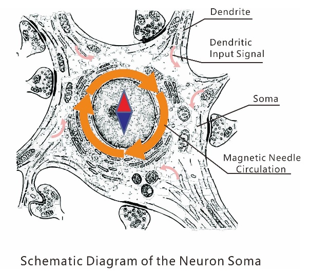
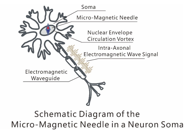 circulating micromagnetic needle. It serves as the minimal information unit participating in brain neural activity, akin to memory units in computers or pixel units in displays. The axon of the neural cell assumes the function of a waveguide for signal transmission, with bidirectional transmission.
3.2 Physical Properties of Micromagnetic Needles
3.2.1 Core Geometric Rule: The magnetic needle is perpendicular to the circulation plane
According to the fundamental laws of electromagnetism, the magnetic field axis generated by a vortex ring current must be perpendicular to the plane of the circulating current. Here, the circulation plane refers to the gyration surface where the neural somata are perpendicular to the axons and intersect with the dendrites. This is a mandatory constraint imposed by electromagnetic geometric laws and forms the core geometric foundation of the micromagnet hypothesis. In the actual anatomical structure of various cortical regions, the axons of pyramidal cells are vertically distributed, and the rotational planes of dendrites are approximately horizontal, which is an inevitable consequence of this physical law.
3.2.2 Sources of Magnetic Moment in Micromagnets
Ionic Circulation:Directed circulation of intracellular ions (e.g., Na⁺, K⁺, Ca²⁺);
Unknown Circulation:Or some unknown structure or electromotive force source forms a vortex circulation driven by signals in dendrites or axons, thereby generating an Ampère magnetic moment;
Permanent Magnetic Particles:Biogenic magnetite (Fe₃O₄) single-domain nanocrystals naturally exist in brain tissue. These particles exhibit typical remanent permanent magnetic properties: under external electromagnetic environmental influences, their magnetic moment orientations can flip, enabling long-term stable retention of inherent magnetic states. This allows information to be stably stored without requiring continuous current or energy input.
3.2.3 Information Encoding Potential of Micromagnets
The direction, strength, and phase of the magnetic moment of the micromagnetic needles can serve as three major dimensions of information encoding: direction corresponds to "information type," strength corresponds to "information weight," and phase corresponds to "information timing." Compared to traditional electrical signal encoding, magnetic encoding offers three key advantages: first, contactless transmission, avoiding synaptic delays; second, strong anti-interference capability, as magnetic field signals are less affected by ion concentration fluctuations; and third, the ability to achieve multi-dimensional parallel encoding, breaking through the single-dimensional limitation of "frequency/phase" in electrical signals.
3.3 Neural Packet Micro-Magnetic Needle Array Networking Mechanism
The orderly arrangement of surface packets forms a "micro-magnetic needle array," whose structural characteristics are closely linked to functionality:
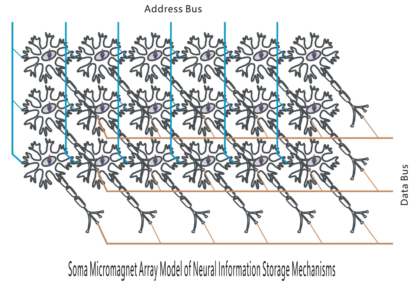
Magnetic Needle Array: All micro-magnetic needles are perpendicular to the cortical surface, ensuring unified magnetic moment direction and forming a natural micro-magnetic needle array with cortical regions as units, providing a geometric foundation for in-phase resonance of neuronal clusters;
Dynamic Networking: The "micro-magnetic needle arrays" in various regions can connect and network through the numerous dendrites of each cell body and surrounding micro-magnetic needle cell bodies, collectively forming a complete scene composition or functional pathway to achieve their corresponding functions.
Density Gradient: The highest density of micro-magnetic needle inclusions (approximately 10⁵–10⁶ per mm³) is found in layers II/III of the gyral protrusions. This high-density arrangement minimizes the near-field magnetic coupling distance between micro-magnetic needles, forming a "functional core area" with strong cooperative oscillations, significantly enhancing local information integration efficiency;
Zonal Independence: The gyral structure divides the array into multiple submodules, each corresponding to a specific function (e.g., vision, hearing). This partitioning mechanism is similar to computer disk partitioning, achieving both functional isolation and reserving pathways for cross-regional axonal wiring;
Information Read/Write: The magnetic moment direction and phase of micro-magnetic needles can be dynamically adjusted through neural activity, enabling information updating and storage.
3.4 Advantages of Micro-Magnetic Needles
Micro-magnetic needle signals are transmitted via "myelin sheath waveguide channels" for low-loss transmission: The insulating properties and cylindrical structure of the myelin sheath form a natural magnetic waveguide, reducing magnetic signal diffusion loss. This ensures synchronous flow of multi-channel magnetic state images, with transmission delays far lower than chemical synapses, and eliminates the need for action potential regeneration along the path.
3.4.1 Memory Storage and Retrieval
- Storage: The topological configurations of magnetic state images remain stable over the long term (permanent magnetism relies on particle lattice stability, while circulating current type depends on ion steady-state regulation), unaffected by the protein's half-life limitation of days to weeks, supporting decades-long memory retention;
- Retrieval: Triggered by "similar cues" — when externally transmitted magnetic state images (e.g., encoded scenes or smells) meet a similarity threshold with stored images, target memories are rapidly activated, perfectly explaining the "associative" characteristic of memory recall.
3.4.2 Sensory Information Decoding and Integration
- Decoding: External sensory stimuli (visual, auditory, etc.) are converted into specific magnetic state images via sensory nerves and rapidly decoded into brain-recognizable information through micro-magnetic needle clusters;
- Integration: Multimodal sensory information (e.g., "seeing an apple + feeling its texture") corresponds to magnetic state images transmitted in parallel via myelinated pathways to the same neuronal clusters. Through image comparison and superposition, integrated unified cognition is formed, explaining the brain's "multisensory fusion" capability.
3.4.3 Low-Power Brain Computation
- Resting Phase: Permanent magnetic state images require zero power to maintain, while circulating current types need minimal energy to stabilize ion gradients, aligning with the brain's low-energy consumption feature during rest periods;
- Operation period: Energy consumption occurs only during the information writing/updating phase. Combined with the high efficiency of parallel magnetic signal transmission, this significantly reduces computational energy consumption, explaining the brain's power balance of "2% body weight proportion, 20% energy consumption."
3.4.4 Cross-Brain Region Information Coordination
The low-loss transmission of myelinated waveguide enables rapid circulation of magnetic state images across brain regions. The endogenous image comparison mechanism ensures precise matching of micro-magnetic needle clusters in different brain regions (such as the cortex, hippocampus, and thalamus), achieving cross-brain region information coordination and explaining the brain's functional characteristic of "global synchronous computation."
3.5 Advantages of Micro-Magnetic Needle Information Encoding
Traditional electrical-chemical encoding is "signal transmission-type" encoding, where information relies on neurotransmitter-driven or current-driven mechanisms. Micro-magnetic needle encoding belongs to field-controlled driving. The core differences between the two are shown in Table 1:
|
Encoding Dimension |
Traditional Electrical-Chemical Encoding |
Micro-Magnetic Needle Magnetic Encoding |
|
Information Carrier |
Action potentials, neurotransmitters |
Magnetic moment direction, strength, phase |
|
Storage Form |
Synaptic strength modification |
Array topological arrangement |
|
Transmission Method |
Synaptic neurotransmitter diffusion |
Magnetic coupling resonance |
|
Transmission delay |
millisecond level (neurotransmitter diffusion) |
microsecond level (contactless coupling) |
|
Anti-interference capability |
Weak (affected by ion concentration) |
Strong (magnetic fields are less susceptible to interference) |
|
Parallel processing capability |
Limited (single-channel transmission) |
Extremely strong (multi-dimensional parallelism) |
4 Empirical Support for the Micromagnetic Needle Hypothesis Theory
4.1 Endogenous Permanent Magnetic Particles and Intracranial Magnetic Oscillations
Numerous authoritative studies have confirmed the presence of biogenic magnetite nanocrystals in the brain. These particles are primarily localized within the cytoplasm of cortical and hippocampal neurons, with their quantity, density, and size fully matching the physical carrier requirements of the micro-magnetic needle hypothesis. Moreover, their distribution regions highly overlap with brain areas associated with memory and information integration. (Kirschvink et al., 1992) further confirmed that these superparamagnetic ferritin particles can form stable magnetic domains, with oscillation frequencies consistent with the neural activity gamma band (40-80 Hz). Modeling studies by (Marrufo-Ramírez et al., 2018) also proposed that neurons can be equivalently described as "magnetic vortex resonators," supporting a same-frequency resonance relay mechanism. These findings provide direct material and magnetic physical foundations for the micro-magnetic needle hypothesis.
4.2 Geometric Compatibility of Inclusions - Dendrites - Axons
Anatomical studies confirm that the somas of cortical pyramidal cells are perpendicular to the cortical surface, with dendrites spreading horizontally and axons initially extending vertically before bending—a structure that is entirely consistent with the geometric principle of "micromagnetic needles perpendicular to the circulation plane" proposed by this hypothesis, serving as the core anatomical support for the hypothesis. Additionally, the histological fact that the sulcal base lacks somas and contains only axons corroborates the design that "grooves serve as wiring channels and ridges as functional working surfaces," aligning closely with the zonal isolation requirements of the micromagnetic needle array.
Why must this arrangement be "nearly strictly" vertical? In biological systems, non-critical structures typically exhibit significant variation, yet the arrangement of cortical pyramidal neurons demonstrates exceptionally high geometric regularity. This strongly suggests that they underpin a core physical function that relies fundamentally on this geometric configuration. The hypothesis posits that this vertical arrangement establishes a unified magnetic field reference coordinate system, enabling orthogonal coupling between micromagnetic needles and dendritic circulation planes while ensuring orderly isolation of axonal transmission pathways. Together, these three elements form the physical foundation of neural information processing.
4.3 Evidence for Vortex Circulation Generation Mechanisms
According to Ampère's circuital law and the Biot-Savart law, the ionic currents within neuronal dendrites generate circular magnetic fields in their surroundings. Nunez & Srinivasan (2006) provided detailed calculations of the macroscopic magnetic field distribution generated by the activity of cortical pyramidal cell populations in their seminal work. They noted that while the current from a single neuron is weak, the synchronized activity of neuronal populations produces magnetic fields detectable by MEG, establishing a theoretical framework for the physical process of "microscopic currents → macroscopic magnetic fields." This hypothesis further proposes that the circular magnetic fields generated by local microcurrent loops in dendrites can couple with the magnetic vortex structures within the cell body, offering a physical basis for the encoding and reading of neural information.
4.4 Axonal Waveguide Transmission Characteristics and Structural Validation
The myelin-axon membrane structure forms a natural dielectric waveguide: the myelin sheath (a low-permittivity lipid layer) acts as the cladding, and the axoplasmic core (high permittivity) serves as the core layer. This configuration enables low-loss electromagnetic signal transmission through total internal reflection (Zarkeshian et al., 2022). Experimental evidence confirms that the electromagnetic signal transmission loss in myelinated axons is only 1/40th of that in unmyelinated axons, meeting the requirements for centimeter-scale transmission across brain regions. When the myelin sheath is damaged, transmission efficiency plummets by 40-fold, further validating the myelin's role in signal confinement.
5.0 Key Arguments Requiring Validation
5.1 Experimental Validation
Using ultra-high-sensitivity magnetic detection technologies (such as superconducting quantum interference devices SQUID, diamond NV center magnetic imaging), directly detect the magnetic moment signals of individual neuronal somas, verify the existence of micromagnetic needles, and establish a quantitative model. Develop a magnetic coupling dynamic model for micromagnetic needle arrays, quantifying the correspondence between magnetic moment direction, strength, phase, and neural activity.
5.2 Validation of Magnetic Moment Flip Triggered by Neural Activity
- Experimental Techniques: Microelectrode arrays (MEA) record neural activity, synchronized with superconducting quantum interference devices (SQUID) to detect local magnetic signals;
- Experimental Content: Induce neurons to generate action potentials (simulating neural activity), observe whether characteristic magnetic signals corresponding to magnetic moment flips occur, and verify the resonant triggering mechanism;
- Expected Results: When neural activity occurs, SQUID detects magnetic signal changes matching the magnetic moment flip of micromagnetic needles, and the energy threshold aligns with theoretical calculations.
5.3 Validation of Myelin Sheath Waveguide Magnetic Transmission
- Experimental Techniques: Prepare in vitro myelinated axon specimens, combined with micromagnetic probes and laser confocal imaging;
- Experimental Content: Apply a simulated micro-magnetic needle magnetic signal to one end of the axon, detect the magnetic signal strength and delay at the other end of the axon, and compare the transmission differences with non-myelinated specimens;
- Expected Result: The magnetic signal transmission loss in myelinated axons is significantly lower than that in the non-myelinated group, and the transmission delay matches the calculated values from waveguide theory.
5.4 Coupling Verification of Cortical Pyramidal Neuron Geometric Arrangement and the Micro-Magnetic Needle Hypothesis
The nearly strict vertical alignment of cortical pyramidal neurons provides critical anatomical structural support for the micro-magnetic needle hypothesis, but the direct relationship between this structure and the function of micro-magnetic needles still requires experimental validation. This section proposes two verifiable core arguments:
5.4.1 Validation of Consistency Between Magnetic Axis Orientation and Soma Long Axis
The hypothesis predicts that endogenous magnetic particles (e.g., Fe₃O₄) within the neuronal soma will align orderly along the long axis of the soma, forming a magnetic axis consistent with the vertical direction of the soma. High-resolution magnetic susceptibility imaging (such as transmission electron microscope magnetic imaging or quantum diamond microscopy) can be used to detect the spatial distribution and orientation of magnetic particles in cortical pyramidal cells, verifying the alignment between their magnetic axis and the soma's long axis. If the particle orientations are random or uncorrelated with the soma axis, the premise of a "unified magnetic field reference coordinate system" in the hypothesis would be invalid.
5.4.2 Validation of Directional Effects of External Magnetic Field Intervention on Cortical Function
The hypothesis posits that the vertical arrangement of cell bodies provides the optimal coupling angle for micromagnetic needles orthogonal to the dendritic current loop plane. By applying directional external magnetic fields (parallel/perpendicular to the cortical surface), differences in neuronal activity, inter-regional synchronization, and related cognitive functions (such as visual topological mapping) under both conditions can be observed. If significant magnetic coupling effects (e.g., enhanced or weakened neural activity synchronization) occur only when the magnetic field direction aligns with the cell body axis, this would provide experimental support for the orthogonal coupling hypothesis. If no significant differences are observed under magnetic field interventions of different directions, the association between geometric arrangement and magnetic coupling would require reevaluation.
6.0 Conclusion
This study proposes and systematically elaborates the theoretical hypothesis that "neuronal somatic micromagnetic needles serve as the fundamental information units of the brain," based on cerebral cortical anatomy, electromagnetic physical principles, and empirical evidence from authoritative literature. This hypothesis makes three core academic contributions: First, overturning traditional paradigms , shifting the focus of neural information processing from "electro-chemical signals" to "magnetic coupling-structural encoding," thereby breaking the century-old theoretical framework of neuroscience. Second, integrating interdisciplinary knowledge , unifying fragmented discoveries from neuroanatomy, electromagnetic physics, biomagnetism, and information science into a cohesive theoretical system for the first time. Third, resolving core challenges , providing verifiable physical mechanisms for "ultimate questions in brain science" such as the origin of consciousness, memory storage, and neural signal transmission.
References
1. Kirschvink, J. L., Kobayashi-Kirschvink, A., & Woodford, B. J. (1992). Magnetite biomineralization in the human brain. Proceedings of the National Academy of Sciences, 89(16), 7683–7687. https://doi.org/10.1073/pnas.89.16.7683
2. Marrufo-Ramírez, J. C., et al. (2018). Magnetic skyrmion dynamics in neuronal systems. arXiv:1809.04563 [physics.bio-ph]. [1809.04563] Finding Higher Order Mutants Using Variational Execution
3. Nunez, P. L., & Srinivasan, R. (2006). Electric fields of the brain: The neurophysics of EEG (2nd ed.). Oxford University Press.
4. Nunez, P. L., Nunez, M. D., & Srinivasan, R. (2019). Multi-scale neural sources of EEG: Genuine, equivalent, and representative. A tutorial review. Brain Topography, 32(2), 193–214.https://www.biorxiv.org/content/10.1101/391318v1.full.pdf
5. Tuszynski, J. A., et al. (2004). Electromagnetic wave propagation in myelinated axons. BioSystems, 77(1-3), 79–91. Redirecting
6. Zarkeshian, P., Kumar, S., Tuszynski, J., Barclay, P., & Simon, C. (2018). Are there optical communication channels in the brain? Frontiers in Bioscience (Landmark Edition), 23(8), 1407–1421. Are there optical communication channels in the brain?
7. Zarkeshian, P., Kergan, T., Ghobadi, R., Nicola, W., & Simon, C. (2022). Photons guided by axons may enable backpropagation-based learning in the brain. Scientific Reports, 12(1), 20720. https://doi.org/10.1038/s41598-022-24871-6

CANN开发者社区旨在汇聚广大开发者,围绕CANN架构重构、算子开发、部署应用优化等核心方向,展开深度交流与思想碰撞,携手共同促进CANN开放生态突破!
更多推荐



 4
4 0
0- 0


 已为社区贡献1条内容

所有评论(0)